The hoof capsule is made up of the same constituent as our hair. This was recently brought home to me at a farrier convention while a horse was being hot shod. The smell of burning hair is too distinct to not recognize. The capsule is made up of tubules that are born and grow down from the coronary papillae, and fused together by an exudate.
The capsule includes the outside wall, buttresses, and bars.
The hoof tubules are longest at the toe, getting shorter and shorter as they round the foot toward the buttress. The wall capsule makes an acute turn at the buttress, and continues back toward the toe-quarter on the opposite side of the foot. The wall tubules continue to shorten toward the natural terminus of the bar, about half way up the frog.
Looking through the hoof capsule…
The outer layer is made up of hard, pigmented tubules. Inside this is a non-pigmented layer of tubules that are softer. And on the inside of this layer, are the insensitive laminae. These three layers make up the wall of the hoof capsule. On the sole plane, the wall is joined to the sole at the white line by the exudate of the terminal papilae of the sensitive lamina on the inner hoof. This exudate is most often referred to as the white line, though it’s yellow.
I’ve tried it. I’ve watched others map a foot while listening to their explanations for why they are doing what they are doing; different styles, explanations and thought processes. None of them were convincing enough to leave me confident that they were pit-fall-free. There were just too many slippery slopes. Too much about mapping is arbitrary. Some still, small voice in me said there was a way to see a hoof that was both more simple, and more clear, and would apply to all hooves.
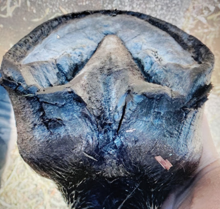
Meanwhile, different aspects of the foot rattled around in my head. All parts of the puzzle. Sorting things out, one by one, bars, buttresses, central sulcuses, when finally…this photo crossed my path as if in answer, and the whole thing clicked.
Seeing this photo was the penny-drop moment for me. One glance at the picture below and I realized that an optimal frog is the hallmark of a healthy, balanced foot. This is a photo of the healthiest, most perfect frog I have ever laid eyes on. Complete with frog curtain.
The state of the frog reveals the state of the hoof. And an optimal frog looks a certain way.
The frog IS the map.
An optimal, and complete frog.
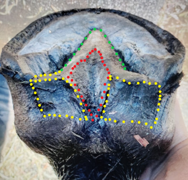
The hallmarks of an optimal frog:
1~ Healthy and not inclined to harbor thrush. Rubbery and firm when cut into.
2~ Straight sides.
3~Natural cleavage into two frog legs, each complete with its own very rubbery frog curtain. The frog curtain is an integral structure of a non compromised frog. It extends directly from the caudal end of each frog leg, over to the heel buttress on the same side. And it grows from the coronet, down to the ground surface. The frog curtain serves to occlude the caudal end of the collateral grove.
Yellow~Frog Curtain. Red~Central Sulcus/Frog Stay Aparatus. Green~Straight Legs
The frog curtain is a structure not often seen. It is the first thing to get compromised out of existence when the trim is off, and one of the last things to redevelop, along with a deeper frog stay, on a foot that is returning to balance and full size. Bracy Clark noted it’s existence on his hoof model circa 1820.
The frog curtain grows down from the periople across the back of the foot and has vertical striations, characteristic of curtains, hence the name. From a liquid dynamics pov, it can be postulated that the frog curtain serves to create a vortex of liquid/mud, when those things are walked or run through, and this resultant vortex flushes out the collateral grooves.
A deep and natural “V” shaped central sulcus is not so much a structure as a void left between fully developed frog legs. A true central sulcus does not need to be cut out , but serves as an indicator of a fully formed frog. The central sulcus opens out of the back of the foot in a ‘V’ when the foot is held in hand. The central sulcus is the underside and external extrapolation of the internal Frog Stay Aparatus, which extends up between the heel bulbs of the digital cushion.
If the frog is not optimal, the foot is not optimal. In an otherwise healthy foot, one not suffering something chronic, like founder or canker, if the frog is not optimal, the capsule is not yet where and how it needs to be. And the foot is most likely smaller than optimal. It is the capsule when trimmed incorrectly, that all to often causes severe compromise in the inner foot, and it’s the inner foot from which the frog and frog curtain grow. Most hooves I see do not have a fully functioning caudal hoof. Frogs are contracted, bars are curved or bent, heels are forward of where they should be, and the caudal foot is smaller than it should be. All of this can be corrected by maintaining proper break-over, and allowing the hind end of the foot to regrow.