Sole Failure, is it the same as Retracted Sole?
When I got Walker (an OTTB) the soles of all four of his feet were slate grey. He never had any retained sole, or chalky sole flaking off; they were always hard and grey. Much like hard plastic. This persisted for the first two to three years.
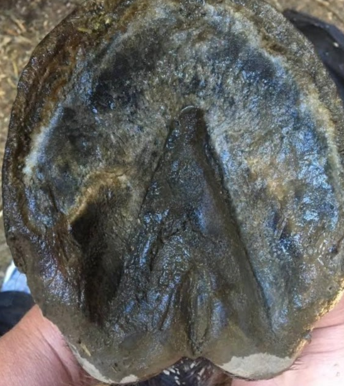
At year 4.5 we were having a nice snowy winter. It was time to trim him and when I picked up the first snow scrubbed foot I was taken aback. Where he’d had only slate grey before, he had a large patch of cream colored sole, with a border of grey. This on his near fore.
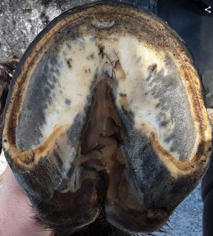
As I checked the rest of his feet, I found his other front also had some cream colored sole with a boarder of grey, though not as much as on his other front. His hinds were still slate grey and hard.
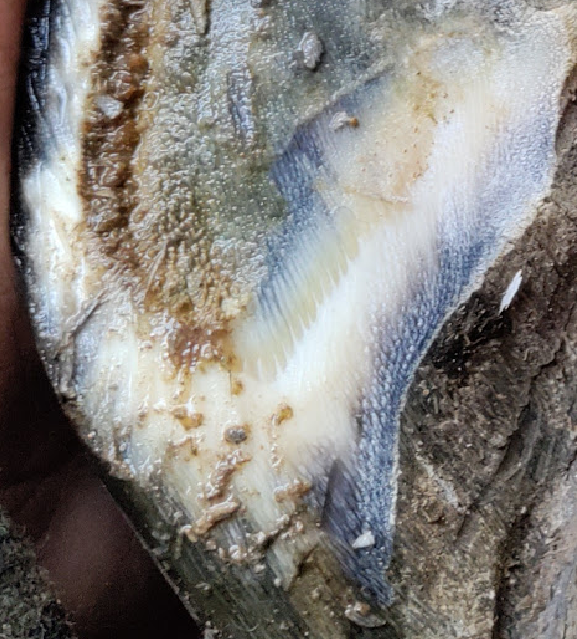
I took slivers of the grey sole material to look at through my stereo microscope. When top lit (light shining on them from the top), they looked as grey as they do to the naked eye. But when I looked at them bottom lit (a light below them shining up through them) it became clear that they were a ruddy color, with more definition to be seen between parts. The ruddy color looks distinctly like dried blood.
It’s my studied opinion that at least on dark colored feet, grey sole is sole that has experienced catastrophic failure, and the grey is the sole tubules, laid over past their optimal angle, devoid of the creamy, inter-tubular matrix, which is present in soles where the tubules of live corium have not been laid over or squeezed shut.
The sole tubules (sensitive and insensitive) lack the stiffness inherent in the capsule tubules. It’s the capsule that provides structural support for the sole tubules. Like how a glass drinking straw holder provides structural support for straws. When capsules go long in the toe and heel buttresses are either folded/laid forward or nonexistent due to having been rasped away, the sole tubules, which lack the stiffness of the capsule tubules, are pressed forward and laid down by ground forces.
Pollitt in his book The Horse’s Hoof has a paragraph on the sole wherein he shared that the vasculature of the sensitive tubules of the sole are arranged in each tubule as a corkscrew. From a fluid dynamics perspective, this reveals each tubule of the sensitive sole corium has the design of a small spring.
It’s reasonable to postulate that as the capsule is born of sensitive papillae that give rise to tubules that are surrounded/encompassed in an inter-tubular matrix , that the sole too is born of it’s sensitive papillae that give rise to sole tubules surrounded by/encompassed in an inter-tubular matrix. The live papillae feed all aspects of the sole including that part which exudes the white waxy substance that makes up healthy, cream colored sole.
When sole papillae are squeezed (when capsules contract), or lay over (when capsules grow or are trimmed below an angle sustainable by the non-ridged tubules, the ability to exude the inter-tubular sole is compromised. When these springs, lacking the required angle of support of the capsule, get laid over, the waxy inter-tubular matrix, lacking the structure provided by the tubules or no longer being created any more by those tubules, ends up wearing away from the sole, leaving it to be replaced by an inter-tubular substance made up of possibly a mixture of blood and lymph, or the same substance that serves as the inter-tubular horn of the capsule.
It’s this waxy inter-tubular sole horn that when present, gives soles their depth.
Laid over sole tubules bound by blood, lymph, or inter-tubular horn of the capsule, have no means of growing depth.
This way of thinking about what is going on in some soles sheds new light on Cheryl Henderson’s ‘beetle experiment’. Cheryl put some cadaver feet in with beetles so they could eat the flesh from the feet, and in some of the hooves she found that not all of the soles of some of the feet were eaten. Some had expanses of harder material left on the sole plane. She postulated that this uneaten sole was actually bar, as the beetles would not eat bar/capsule. But what the beetles would not eat was not bar, but sole that had experienced failure and had inter-tubular sole wax replaced by blood and lymph (or perhaps, by the same substance that binds the capsule tubules together) It seems to me that the beetles liked the waxy sole, but were disinclined to eat the failed sole.
I believe it’s a catastrophic laying over of sole tubules that causes the phenomenon of ‘retracted sole’. The tubules lay down from the back to the front of the hoof, and when that laying down reaches the capsule wall the tubules stack up against it, creating the characteristic ramp to the distal/ground edge of the capsule. And when soles have spots of black/grey, that is from the live sole corium being squeezed forward by circumstances (trimming or keep) that cause the capsule to shrink forward.
I’ve reason to believe that the hard sole that develops in failed sole is fortified by the same, or some of the same substance that glues the outer pigmented capsule tubules together. In dark footed horses, a failed sole is grey, and in light soled horses I’ve seen it to be almost orange. So it seems as if this harder glue is in part pigmented by the same substance that pigments the outer capsule. Thus far, the only truly waxy sole I’ve seen is cream colored.
One last thing I noticed was that when I first started the process of bringing back Walker’s toes when his soles were grey, and I rasped into sole at the toe, this grey sole seemed to have moisture in it, again, I wonder about lymph.